Spinal Navigation Systems: How Technology Improves Outcomes
In the realm of modern medicine, few fields have seen a transformation as radical as spinal surgery. Traditionally, spinal procedures relied heavily on a surgeon’s deep knowledge of anatomy, tactile feedback, and intermittent 2D X-ray images (fluoroscopy). While effective, this “freehand” method left a margin for error, particularly in complex deformities.
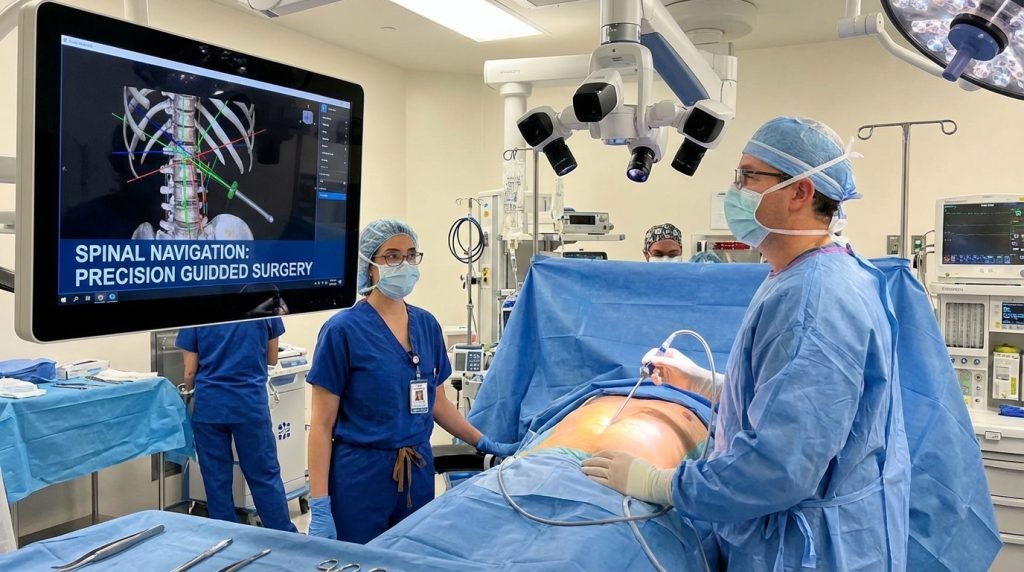
Today, Spinal Navigation Systems have emerged as the “GPS of the operating room.” By integrating advanced imaging with real-time tracking, these systems are redefining surgical precision and significantly improving patient outcomes.
What are Spinal Navigation Systems?
At its core, a spinal navigation system is a computer-assisted technology that allows surgeons to visualize the patient’s anatomy in three dimensions during a procedure. It functions similarly to a satellite navigation system in a car; just as a GPS shows a vehicle’s position on a digital map, spinal navigation shows the exact location of surgical instruments relative to the patient’s spine.
The Core Components
- Imaging Input: Pre-operative CT/MRI scans or intraoperative 3D scans (like the O-arm).
- Tracking Camera: An infrared or optical sensor that monitors the position of instruments.
- Reference Frames: Small markers attached to the patient’s bone to provide a constant point of orientation.
- The Computer Console: The “brain” that fuses the images and tracking data into a real-time 3D display.
The Evolution: From “Freehand” to 3D Guidance
To understand the impact of navigation, one must look at the traditional approach. In conventional spine surgery, surgeons used fluoroscopy—essentially a series of still X-rays taken during the operation. This provided only a flat, two-dimensional view of a complex, three-dimensional structure.
The shift toward Image-Guided Spine Surgery (IGSS) began in the late 1990s and has accelerated with the advent of high-speed processing and robotic integration. We have moved from:
- Tactile Feedback: Relying on the “feel” of the bone.
- 2D Fluoroscopy: Frequent X-rays that increased radiation exposure.
- 3D Navigation: Real-time, sub-millimeter accuracy that allows for proactive rather than reactive surgery.
How Spinal Navigation Improves Surgical Outcomes
The primary goal of any surgical innovation is to improve the patient’s quality of life while reducing risk. Spinal navigation achieves this through several critical mechanisms.
1. Unmatched Accuracy in Pedicle Screw Placement
One of the most common spinal procedures is fusion, which often requires the placement of pedicle screws to stabilize the vertebrae. In the freehand technique, screw malposition rates can vary. However, studies consistently show that spinal navigation systems increase screw placement accuracy to over 95-98%.
By seeing the internal trajectory in real-time, surgeons can avoid critical structures like the spinal cord, nerve roots, and major blood vessels.
2. Reduction in Radiation Exposure
In traditional surgery, the surgical team and the patient are exposed to repeated bursts of X-ray radiation to confirm instrument placement. Computer-assisted spine surgery allows for “virtual fluoroscopy.” Once the initial 3D scan is taken and registered, the surgeon can navigate without needing constant new X-rays, drastically reducing the lifetime radiation dose for both the patient and the medical staff.
3. Facilitating Minimally Invasive Spine Surgery (MISS)
The “holy grail” of modern orthopedics is achieving the same results through smaller incisions. Traditionally, surgeons needed large incisions to “see” the anatomy.
With spinal navigation, the computer provides the visualization. This allows for:
- Smaller incisions.
- Less muscle stripping and tissue trauma.
- Reduced blood loss during surgery.
- Faster recovery times and shorter hospital stays.
4. Enhanced Decision-Making in Complex Deformities
For patients with severe scoliosis or complex tumors, the anatomy is often distorted. Navigation systems allow surgeons to map out the most dangerous areas before making a single cut. The ability to visualize the “hidden” side of a bone or the exact borders of a tumor ensures a more complete resection and a safer reconstruction.
Key Technologies Leading the Market
Several industry leaders have developed proprietary systems that are now standard in high-performing neurosurgical centers:
- Medtronic StealthStation™ & O-arm™: Perhaps the most well-known duo, the O-arm provides intraoperative 3D imaging that automatically syncs with the StealthStation navigation software.
- Stryker NAV3i®: Known for its high-resolution optical tracking and “plug-and-play” integration with various surgical instruments.
- Globus Medical ExcelsiusGPS®: A leader in the transition toward robotic-assisted navigation, combining a rigid robotic arm with navigation software for even greater stability.
- Brainlab Curve™: Focuses on high-end image manipulation and data sharing, allowing for integrated digital ORs.
Challenges and Considerations
Despite the clear benefits, the adoption of spinal navigation is not without hurdles.
- The Learning Curve: Surgeons must undergo specialized training to become proficient. There is a transition period where surgery may actually take longer as the team learns to calibrate the equipment.
- Initial Cost: These systems represent a multi-million dollar investment for hospitals. However, proponents argue that the reduction in re-operation rates (due to misplaced screws) and shorter hospital stays provide a significant long-term Return on Investment (ROI).
- Registration Errors: If the reference frame moves or the patient shifts, the “map” becomes inaccurate. Surgeons must remain vigilant and “trust but verify” the digital feedback against their anatomical knowledge.
The Future: AI, Augmented Reality (AR), and Robotics
The next frontier for spinal navigation is the integration of Artificial Intelligence (AI) and Augmented Reality (AR).
- AI-Driven Planning: In the future, software will be able to suggest the optimal screw size and trajectory based on thousands of previous successful cases, acting as a digital consultant to the surgeon.
- AR Headsets: Instead of looking away from the patient at a computer screen, surgeons are beginning to use headsets (like the xvision system) that overlay the 3D spinal anatomy directly onto the patient’s back. This creates a “X-ray vision” effect, keeping the surgeon’s eyes on the operative field at all times.
- Robotic Surgery: While navigation tells the surgeon where to go, robotics ensures they stay on that path, eliminating the risk of hand tremors or tool slippage.

