
When someone comes in with sudden, electric-shock pain on one side of the face, the first question is simple: what is irritating the nerve? In many cases, the answer is a small blood vessel pressing against it. And the best way to look for that vessel, without opening anything up, is an MRI.
An MRI for trigeminal neuralgia is not a routine scan. It’s a carefully planned study, and the findings can decide whether microvascular decompression (MVD) is even worth considering. Let’s walk through what the scan looks for and what it can and can’t tell us.
Why the MRI Matters So Much
Trigeminal neuralgia is mostly diagnosed from your story: where the pain is, how it feels, what sets it off. No scan can “see” pain. But the MRI answers a different question: is there a physical cause we can treat?
It helps in three ways:
- It shows whether a blood vessel is pressing on the trigeminal nerve.
- It rules out other causes, such as a tumour or a cyst near the nerve.
- It helps pick up conditions like multiple sclerosis, which can also cause facial pain.
For anyone being considered for MVD, this information is essential. The surgery works by moving a vessel away from the nerve, so we need to know, as clearly as possible, that such a vessel exists.
What Is Being Looked At
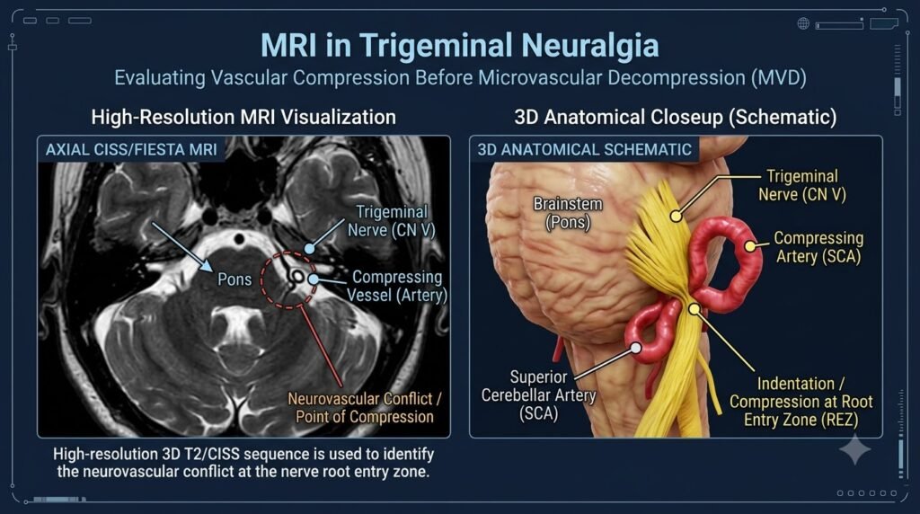
The trigeminal nerve leaves the brainstem and travels toward the face. The most sensitive spot is where it first leaves the brainstem, called the root entry zone. This is where a looping artery or vein most often presses on it.
The artery most commonly involved is the superior cerebellar artery, though other vessels can be responsible too. A good MRI shows the nerve, the nearby vessels, and the small gap between them, all in fine detail.
The Type of MRI Makes a Difference
A basic brain MRI can easily miss a tiny vessel touching a thin nerve. That’s why doctors ask for special high-resolution sequences focused on the nerve. Two are commonly used:
- Heavy T2-weighted sequences (often known by names like CISS or FIESTA), which show the nerve clearly against the fluid around it.
- MR angiography (MRA), which highlights the blood vessels so the artery can be told apart from the nerve.
Doctors often combine these views, so they can follow the vessel and nerve in three dimensions. Many centres prefer a 3 Tesla scanner for sharper images, though a good study on a 1.5 Tesla machine can also be useful.
Sometimes contrast dye is added, mainly to rule out tumours or inflammation. Whether it’s needed depends on your case.
Contact Is Not the Same as Compression
This is the part many patients find surprising. Seeing a blood vessel near the nerve does not automatically mean it’s the cause of the pain. Plenty of people have vessels touching the trigeminal nerve and never feel a thing.
What doctors look for is true compression, meaning signs like:
- The nerve looks flattened, bent, or pushed out of its normal position.
- A groove or indentation is visible on the nerve.
- The nerve on the painful side looks thinner than the one on the other side.
- The vessel sits right at the root entry zone, on the same side as the pain.
The scan has to be read alongside your symptoms. A finding on the MRI only matters if it matches what you feel.
What the MRI Cannot Tell Us
It’s worth being honest about the limits. An MRI isn’t perfect.
- It can occasionally miss a small compressing vessel.
- It can suggest compression when the surgeon finds something different during the operation.
- A normal-looking MRI doesn’t always rule out trigeminal neuralgia.
So a scan that shows no clear compression doesn’t automatically close the door on surgery, and a scan that shows contact doesn’t automatically open it. The decision comes from putting together your symptoms, your response to medicines, and your imaging. That’s why an experienced neurosurgeon, not just a radiology report, should review the pictures.
What to Expect on the Day of the Scan
The scan is painless. You’ll lie still on a table that slides into the machine, and it makes loud knocking sounds, so you’ll be given earplugs or headphones. It usually takes around 30 to 45 minutes.
A few practical tips:
- Tell the team if you have a pacemaker, metal implants, or a fear of closed spaces.
- If you’re given contrast, let them know about any kidney problems or allergies.
- Try to stay as still as you can, since movement blurs the fine details.
After the Scan
Once the images are ready, your doctor will go through them with you. If compression is clear and your symptoms fit, MVD may be a strong option. If the findings are unclear, other treatments may be discussed, or further tests may be needed. Either way, you’ll know where you stand.
For patients considering MVD surgery for facial pain, the MRI findings are an important part of understanding whether vascular compression may be contributing to their symptoms and whether MVD is appropriate.
Frequently Asked Questions
Can an MRI confirm trigeminal neuralgia?
Not on its own. The diagnosis mainly depends on your symptoms. The MRI helps find the cause and rule out other conditions.
Is every blood vessel near the nerve a problem?
No. Contact alone is common and often harmless. Doctors look for actual compression, such as a displaced or flattened nerve.
Do I need contrast for the MRI?
Not always. It’s used when your doctor wants to check for tumours or inflammation.
What if my MRI looks normal?
You may still have trigeminal neuralgia. A normal scan means no obvious compression was seen, and your doctor will decide the next step based on your overall picture.
Is the MRI painful or risky?
It’s painless and doesn’t use radiation. The main concerns are metal implants and claustrophobia, so share these beforehand.
Can I get MVD only if the MRI shows compression?
Clear compression makes you a stronger candidate, but the final decision also depends on your symptoms, medicines, and general health.
Final Thoughts
An MRI is one of the most useful tools in planning treatment for trigeminal neuralgia. It helps show whether a blood vessel is truly pressing on the nerve, rules out other causes, and guides the decision about MVD. But the scan is only one piece. Your story, your medicines, and an experienced specialist’s judgement matter just as much.
If you are looking for the best neurosurgeon for MVD in India, an experienced specialist can review your MRI, symptoms and treatment history to help determine whether MVD is the right option for you.

